Recent advancements in microscopy have led to a breakthrough in the field of life sciences. A team of researchers at HHMI’s Janelia Research Campus have found a way to adapt techniques from astronomy to improve the clarity and sharpness of microscopy images, providing biologists with a more efficient and cost-effective method for imaging biological samples. The findings, published in the journal Optica, reveal a new approach that could revolutionize the way biologists study cells and tissues.
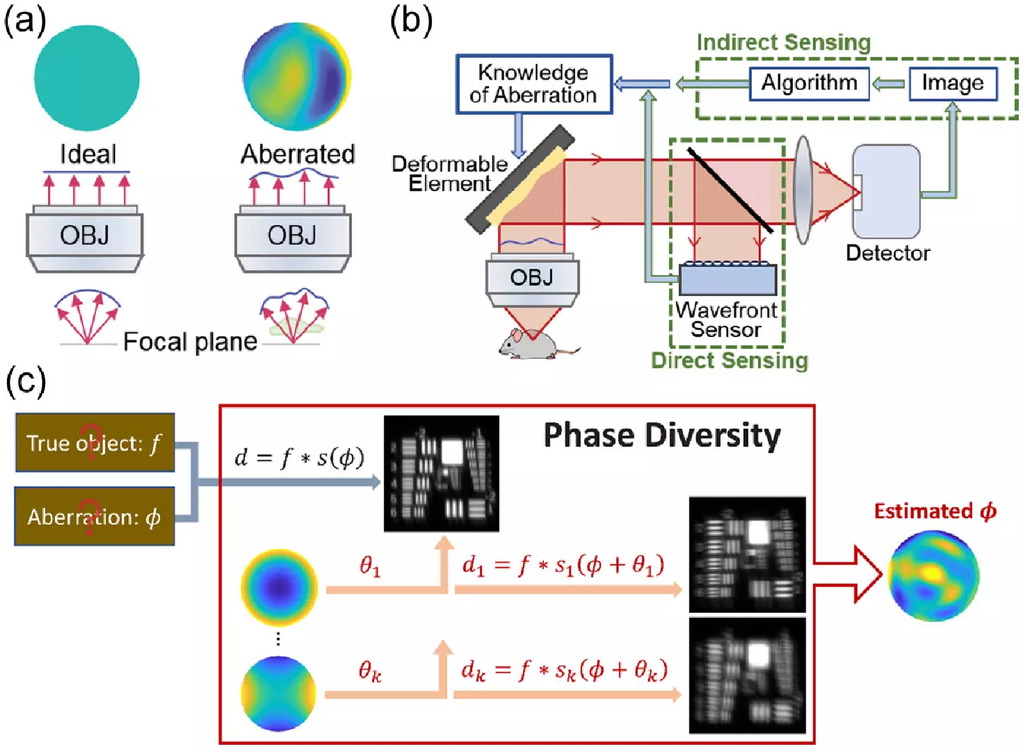
Microscopists face the challenge of capturing clear and sharp images of thick biological samples that distort light and create aberrations. Traditional methods for correcting these distortions, such as adaptive optics, are complex, expensive, and time-consuming, making them inaccessible to many labs. The team at Janelia Research Campus sought to address these limitations by exploring phase diversity techniques previously used in astronomy.
Phase diversity methods involve adding images with known aberrations to a blurry image with unknown aberrations, providing additional information to unblur the original image. Unlike other adaptive optics techniques, phase diversity does not require major changes to an imaging system, making it a more practical solution for microscopy. The team modified an astronomy algorithm for microscopy, built a microscope with a deformable mirror and additional lenses, and enhanced the software for phase diversity correction.
The team conducted simulations to validate their new method and demonstrated significant improvements in calibration speed and image clarity. By sensing and correcting randomly generated aberrations, the researchers were able to obtain clearer images of fluorescent beads and fixed cells. The next phase of the research will involve testing the method on real-world samples, including living cells and tissues, to further refine its applicability to complex microscopes.
The researchers aim to automate and simplify the method for broader use in labs. By providing a faster and more cost-effective alternative to current techniques, the new method could potentially democratize access to adaptive optics in microscopy. This development holds promise for biologists seeking to enhance their ability to study cellular structures and processes with better clarity and precision.
Overall, the innovative approach of applying phase diversity techniques from astronomy to microscopy represents a significant advancement in the field of life sciences. With further testing and refinement, this method could open new doors for researchers to explore the intricate details of biological samples with unprecedented clarity and accuracy.


Leave a Reply