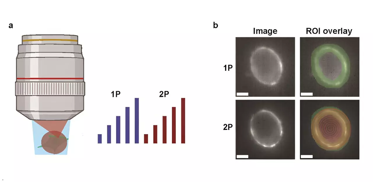
The intricate workings of neural circuits within the brain have long intrigued scientists striving to decode the mechanisms of neuronal communication and information processing. Foremost in this endeavor are genetically encoded voltage indicators (GEVIs), which facilitate the visualization of electrical activity in real-time. These tools are pivotal for developing our understanding of neural dynamics. Nonetheless, a significant debate persists regarding the efficacy of two primary imaging techniques: one-photon (1P) and two-photon (2P) voltage imaging.
A recent investigation conducted by a team at Harvard University provides a deeper insight into the comparative advantages and disadvantages of 1P and 2P voltage imaging methodologies. As highlighted in their publication in *Neurophotonics*, the researchers meticulously contrasted these techniques, taking into account the optical constraints and biophysical limitations intrinsic to each. Their research not only assessed the brightness and voltage sensitivity of various GEVIs in different settings but also examined the degradation of fluorescence with increasing depth in the mouse brain—a pivotal aspect for effective in vivo imaging.
The implementation of a quantitative model enabled the team to predict the quantity of measurable neurons based on the characteristics of the indicators, the parameters of imaging, and targeted signal-to-noise ratio (SNR). This analytical approach allowed for a clearer understanding of the impact of technological improvements in imaging and sensor design on voltage measurement efficacy.
One of the pivotal revelations of the study is that 2P voltage imaging demands an astonishingly higher illumination power, approximately 10,000 times that required for 1P imaging, to achieve analogous photon count rates. This immense power requirement raises critical concerns regarding potential tissue damage and the introduction of shot noise, complicating the application of this method for in vivo studies. For instance, the use of a specific 2P indicator, JEDI-2P, in mouse cortex assessments targets SNR values of around 10, but with limitations on laser power and bandwidth, it can feasibly capture activity from only about a dozen neurons at depths exceeding 300 micrometers.
In light of the findings from this comparative study, the researchers concluded that the modest voltage sensitivity of current GEVIs, in conjunction with stringent photon-count prerequisites, renders 2P voltage imaging particularly challenging in live specimens. To enable the capture of high SNR from a greater number of neurons at significant depths, the field must either enhance existing 2P techniques or pioneer new imaging strategies altogether.
This investigation serves as a clarion call for continued innovation in imaging technologies, emphasizing the inherent trade-offs between alternative imaging modalities. As the scientific community seeks to deepen its comprehension of neural circuitry through advanced imaging, the insights from this research are poised to guide future technological developments. Embracing these challenges and fostering innovative solutions will ultimately illuminate the enigmatic nature of the brain’s electrical activities, bringing groundbreaking advancements to neuroscience.
Leave a Reply