In the rapidly advancing field of biotechnology, the demand for innovative methodologies to observe biomolecules within living cells is paramount. The National Institute of Standards and Technology (NIST) has made significant strides in this area by unveiling a novel technique that employs infrared (IR) light to produce clear images of biomolecules inside cells. This development is poised to accelerate advancements in drug therapies and biomanufacturing, addressing a critical gap in current imaging technologies.
Historically, imaging biomolecules within cells has been impeded by the overwhelming presence of water, which absorbs infrared radiation. This absorption often obscures valuable insights into the biomolecular landscape of living cells. As a result, researchers have struggled to accurately measure vital components such as proteins and nucleic acids, which play crucial roles in cellular function and health. The traditional methods often fell short, requiring extensive resources and complex setups, such as synchrotron facilities, to obtain relevant data.
To illustrate the dilemma, one might liken the situation to watching an aircraft through a glare of sunlight—it’s challenging without specialized filters that enhance visibility. The new method introduced by NIST focuses on overcoming this optical masking effect, thereby revealing the hidden dynamics of biomolecular interactions in live cells.
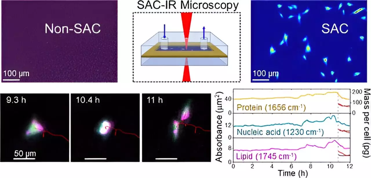
At the heart of NIST’s innovation is a patented technique known as solvent absorption compensation (SAC). Developed by chemist Young Jong Lee, this method employs an advanced optical system that effectively compensates for water’s absorption characteristics in IR measurements. By distinguishing water’s influence from that of biomolecules, researchers can now unveil the infrared absorption spectra of proteins and other key molecules.
Utilizing a custom-built IR laser microscope, Lee and his team conducted 12-hour imaging sessions on fibroblast cells, which are essential for the formation of connective tissue. Over this duration, they successfully tracked changes in the cellular composition during various stages of the cell cycle, capturing a comprehensive snapshot of biomolecular dynamics that would have been previously unattainable.
One of the standout features of the SAC-IR method is its label-free approach. Conventional imaging techniques often rely on dyes and fluorescent markers that can harm cells and produce inconsistent results. In contrast, SAC-IR facilitates the measurement of absolute quantities of proteins, nucleic acids, lipids, and carbohydrates without these intrusive labels. This capability not only preserves the integrity of living cells but also paves the way for standardized measurement protocols across different research laboratories.
This aspect is particularly critical in fields such as cancer cell therapy, where evaluating the safety and efficacy of modified immune cells is essential. With SAC-IR, researchers can garner invaluable insights into the biomolecular changes taking place in these cells, ultimately leading to more effective therapeutic strategies.
The implications of the SAC-IR technique extend far beyond mere imaging; it has the potential to revolutionize drug development. By enabling scientists to assess the concentration of biomolecules at an individual cell level, the method could facilitate high-throughput screening of drug candidates, assessing their potency and the cellular responses to various treatments.
Furthermore, as researchers continue to refine the technique, there is hope for improved accuracy in measuring additional biomolecules, including DNA and RNA. Such progress could yield profound insights into fundamental biological processes, including determining the viability of cells—essential information for applications ranging from regenerative medicine to biopreservation.
For instance, many cell types are stored in frozen states and may lose viability upon thawing. NIST’s new measurement capabilities could lead to better methodologies for managing these processes by analyzing the biomolecular signatures that signify a cell’s health status upon revival.
The advancements brought forth by the NIST researchers represent a pivotal shift in the methodology of biomolecular imaging. By addressing the long-standing challenges posed by water absorption in infrared microscopy, the SAC-IR technique opens up new avenues for research and application in biotechnology. As the scientific community embraces this innovative approach, the potential for developing lifesaving therapies grows exponentially, heralding a transformative era in the way we visualize and understand the complexities of living cells. Through ongoing research and development, we can anticipate more breakthroughs that will strengthen our grasp of cellular processes and enhance the efficacy of therapeutic interventions.


Leave a Reply