Researchers in various scientific fields have been striving to develop innovative techniques for imaging weakly scattering phase objects, such as cells. Chemical stains and fluorescent tags have traditionally been used to enhance image contrast, but these methods often involve complex and potentially toxic sample preparation steps. However, a breakthrough in the form of quantitative phase imaging (QPI) has provided a label-free solution that offers non-invasive, high-resolution imaging of transparent specimens without the need for external tags or reagents.
Despite the advantages of QPI, traditional systems have their limitations. They can be resource-intensive and slow due to the reliance on digital image reconstruction and phase retrieval algorithms. Moreover, most QPI approaches do not account for the presence of random scattering media commonly found in biological tissue. These limitations have hindered the widespread adoption of QPI in various scientific disciplines.
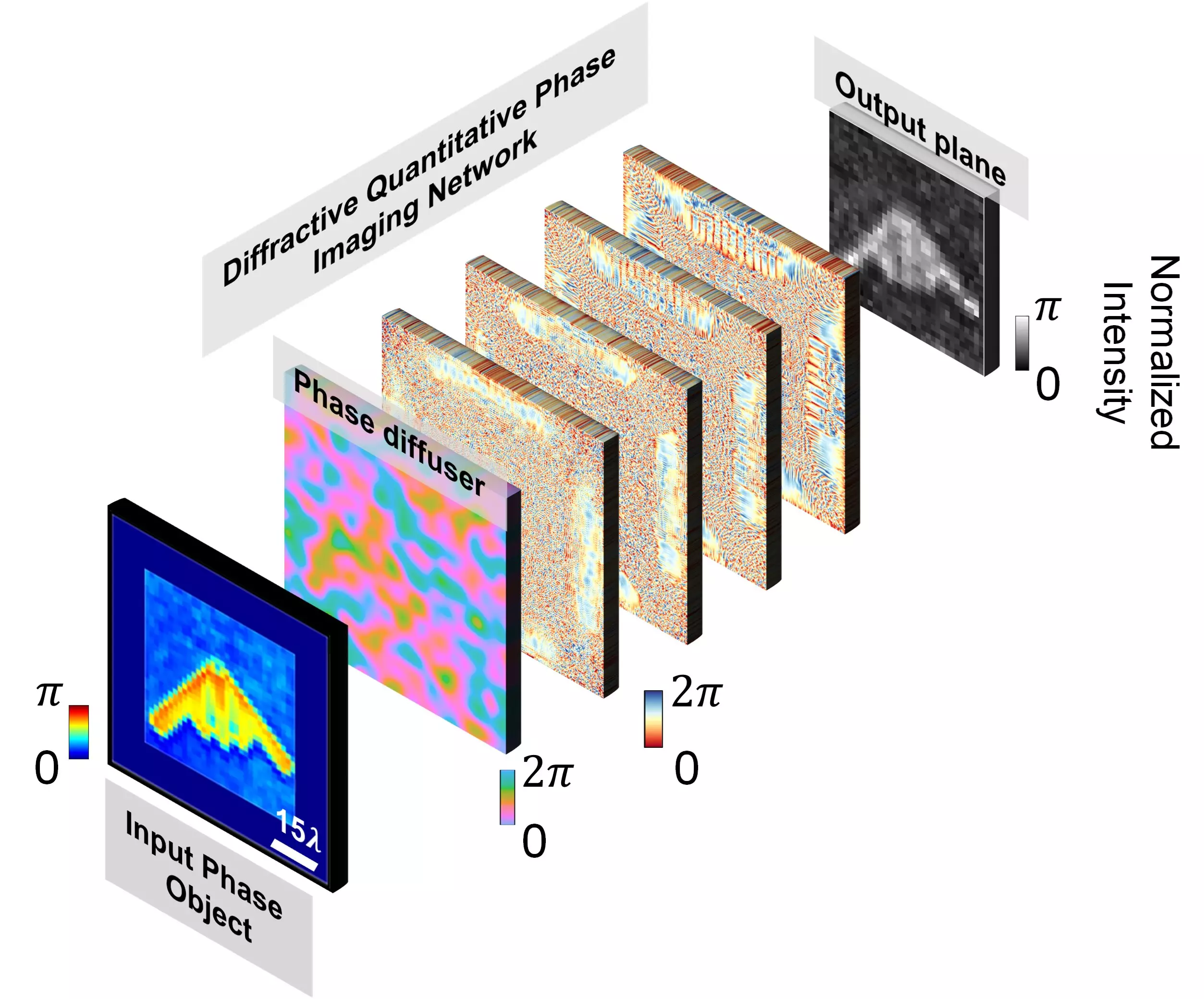
Professor Aydogan Ozcan and his research team from the University of California, Los Angeles (UCLA), have recently presented a groundbreaking methodology for quantitative phase imaging of objects completely covered by random, unknown phase diffusers. The researchers developed a diffractive optical network comprising multiple transmissive layers that were optimized using deep learning techniques. Notably, this innovative diffractive system spanned approximately 70 times the illumination wavelength.
During the training phase, the research team utilized randomly generated phase diffusers to build resilience against phase perturbations caused by unknown diffusers. Upon completion of the training, the resulting diffractive layers were capable of performing all-optical phase recovery and quantitative phase imaging of objects hidden by unknown random diffusers. The team successfully demonstrated the effectiveness of the QPI diffractive network in imaging new objects through previously unseen random phase diffusers using numerical simulations.
To further optimize the performance of the QPI diffractive network, the researchers investigated the influence of the number of spatially-structured diffractive layers and the trade-off between image quality and output energy efficiency. Deeper diffractive optical networks were found to generally outperform shallower designs. Importantly, the QPI diffractive network can be easily scaled to operate at different parts of the electromagnetic spectrum without the need for redesigning or retraining its layers.
The all-optical computing framework developed by the UCLA research team offers several advantages, including low power consumption, high frame rate, and a compact size. The researchers envision the integration of their QPI diffractive designs onto image sensor chips, such as CMOS/CCD imagers. This integration would transform a conventional optical microscope into a diffractive QPI microscope capable of on-chip phase recovery and image reconstruction through light diffraction within passive structured layers.
This revolutionary approach to quantitative phase imaging has the potential to revolutionize various fields, particularly biomedical sciences. The label-free nature of the technique eliminates the need for toxic or destructive sample preparation steps, making it safer and more efficient. Additionally, the ability to image objects hidden by unknown random diffusers opens up new possibilities for studying biological tissue and other complex specimens. The scalability of the QPI diffractive network further enhances its versatility and applicability across different parts of the electromagnetic spectrum.
Professor Aydogan Ozcan and his team at UCLA have introduced a game-changing methodology for quantitative phase imaging. By combining deep learning techniques with a diffractive optical network, they have overcome the limitations of traditional QPI systems and paved the way for high-resolution imaging of weakly scattering phase objects in a label-free and non-invasive manner. The potential applications of this technology are vast, and its integration into image sensor chips holds great promise for the future of microscopy. With this groundbreaking development, the scientific community can look forward to new insights and advancements in various fields.


Leave a Reply