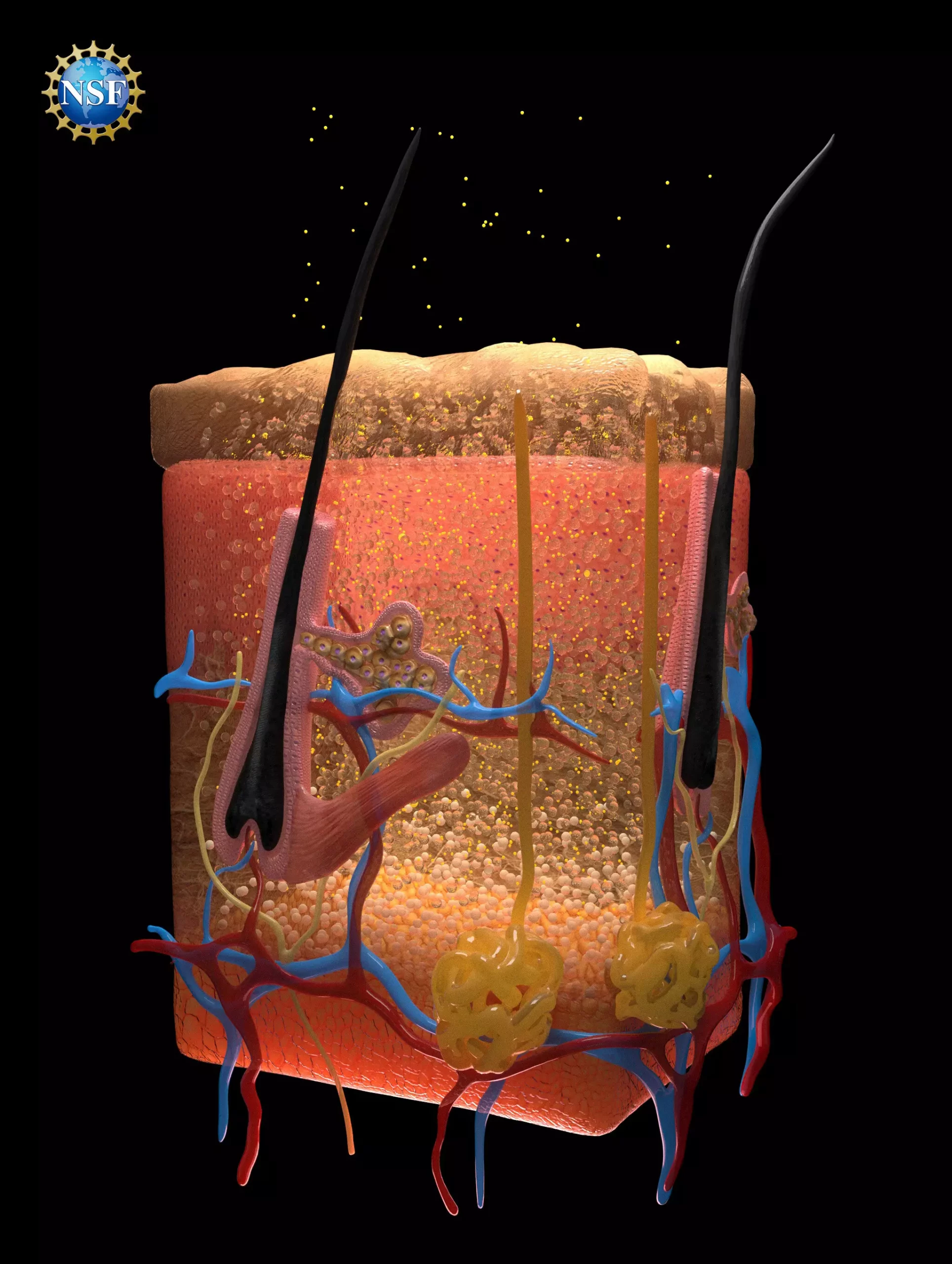
Recent breakthroughs in medical imaging have highlighted the potential of creating transparent biological tissues, facilitating better visibility of internal organs. Researchers at Stanford University have devised a fascinating method that involves rendering tissue transparent through the use of food-safe dyes. This innovative technique, published in the prestigious journal *Science*, opens the door to significant advancements in medical diagnostics, offering new avenues for locating injuries, monitoring digestive disorders, and identifying cancerous cells.
The core of this transparency technique revolves around the modification of light scattering properties in biological tissues. Light scattering is a phenomenon that occurs due to the varied refractive indices of materials within the body, such as fats, fluids, and proteins. When light passes through these materials, it changes direction, which usually results in the opacity we observe in biological tissues. The research team recognized that to achieve transparency, they needed to synchronize the refractive indices of these diverse materials.
They employed a sophisticated understanding of optics to create a model predicting how dyes interact with biological tissues. Remarkably, they discovered that the right dyes could not only absorb certain wavelengths of light but could also facilitate a more uniform passage of light through biological materials by adjusting their refractive indices. This dual functionality proved essential for the transparency effect.
Among the dyes analyzed, tartrazine — commonly known as FD & C Yellow 5 — emerged as a leading candidate due to its ability to match refractive indices effectively. When dissolved in water and applied to tissues, tartrazine demonstrated an exceptional capability to minimize light scattering, rendering the selected tissues transparent. Initial experiments with thin samples of chicken breast showcased this process convincingly; as the concentration of the tartrazine solution increased, transparency followed suit.
Furthermore, the application was expanded from in vitro samples to live animals, where tartrazine was gently rubbed onto the skin of mice. The results were astounding. The dye effectively highlighted previously unseen structures, such as blood vessels beneath the skin, and made internal processes like heartbeats and intestinal contractions observable. Remarkably, once the dye was removed, tissues returned to their normal state without any lasting effects, indicating the reversible nature of this technique.
The repercussions of this discovery for medical practice are profound. This technique could potentially enhance procedures involving blood draws by providing clearer visibility of veins. Moreover, the method could simplify processes reliant on laser treatments — particularly for issues like tattoo removal or in the field of oncology, where lasers are used to eradicate malignant cells. By improving light penetration depth, it could enable treatments targeting deeper tissues, transforming therapeutic approaches to cancer treatment.
Researchers speculate that further refinement of this technique through the injection of dye could yield even more detailed insights into internal biological processes in living organisms. The prospect of using optical techniques to visualize the internal workings of the body is revolutionary and could significantly enhance diagnostic capabilities.
Scientific Background and Collaborative Efforts
This groundbreaking research stems from an investigation into the interaction of microwave radiation with biological tissues, which serendipitously led to insights applicable to visible light. An exploration of classic optics literature provided crucial mathematical frameworks, including the Kramers-Kronig relations, that guided the researchers in their experimentation with dyes.
The study saw robust collaboration among 21 researchers, emphasizing the importance of diverse expertise in scientific innovation. One of the standout instruments used during the research was an ellipsometer, a tool traditionally utilized in the semiconductor industry that proved its worth in studying the optical properties of target dyes. Such cross-disciplinary applications exemplify the unexpected paths that lead to significant advancements in various fields of science.
The research team’s findings delineate a new frontier in medical imaging and diagnostics. By merging principles of physics with practical applications in medicine, they have not only demonstrated a method for achieving tissue transparency but have also set the stage for a revolutionary approach to understanding and diagnosing complex biological processes.
As research continues, we anticipate discovering new dyes and methods for further enhancing light interaction with biological tissues. The hope is to see this technique applied in clinical settings, paving the way for heightened diagnostic accuracy and improved patient outcomes. The fusion of theoretical understanding with innovative practice holds tremendous promise, and the world awaits the next leap forward in medical imaging technology.


Leave a Reply