The world of microscopy is an essential tool for scientists and researchers to delve into the microscopic realm and explore the inner workings of cells, viruses, proteins, and molecules. While modern microscopes have provided us with incredible insights, they are not without their limitations. Traditional microscopy techniques, such as super-resolution fluorescent and electron microscopy, have constraints that hinder their effectiveness in studying live cell samples. Super-resolution fluorescent microscopy requires labeling specimens with fluorescence, which can be toxic and lead to sample bleaching with extended light exposure. On the other hand, electron microscopy provides outstanding details but requires samples to be placed in a vacuum, making it impossible to study live samples. These limitations have driven the search for more advanced and non-invasive imaging techniques.
Mid-infrared microscopy has long been considered a promising imaging technique due to its ability to provide both chemical and structural information about live cells without the need for labeling or damaging the samples. However, its application in biological research has been limited by its relatively low resolution capability, typically around 3 microns. In a groundbreaking development, a team of researchers at the University of Tokyo has pushed the boundaries of mid-infrared microscopy by achieving a spatial resolution of 120 nanometers, a remarkable 30-fold improvement over conventional mid-infrared microscopes. This advancement opens up new possibilities for studying infectious diseases and developing more accurate imaging techniques in the future.
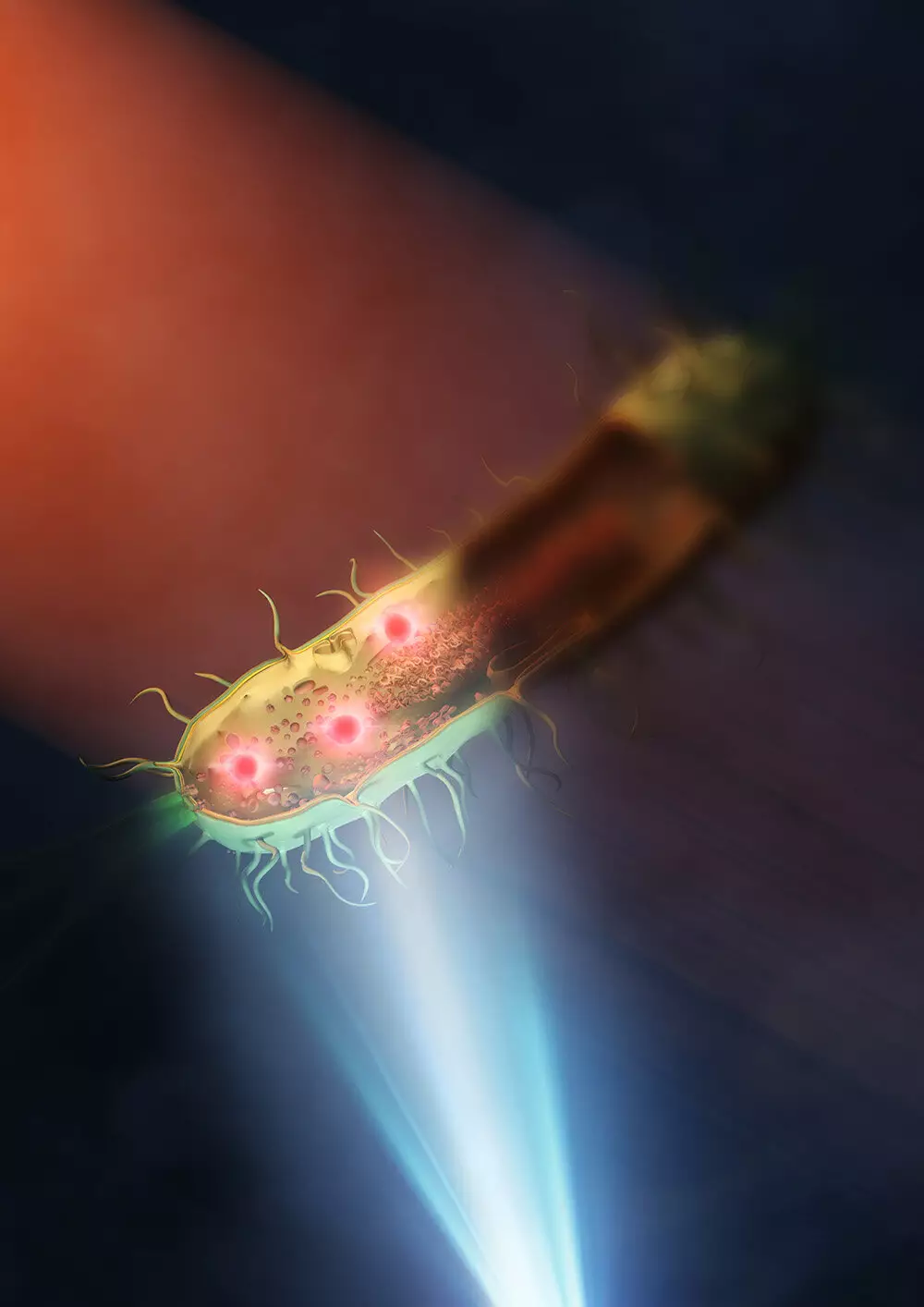
The team at the University of Tokyo utilized a synthetic aperture technique, combining images taken from different illuminated angles to create a clearer overall picture. By placing the live bacteria samples, E. coli and Rhodococcus jostii RHA1, on a silicon plate that reflected visible light and transmitted infrared light, they were able to eliminate the issue of lens absorption of mid-infrared light. This innovative approach allowed the researchers to use a single lens to better illuminate the samples with mid-infrared light and obtain highly detailed images of the intracellular structures of bacteria. Professor Takuro Ideguchi from the Institute for Photon Science and Technology at the University of Tokyo expressed their surprise at the clarity of the images and highlighted the potential of this technology in studying antimicrobial resistance and other global health challenges.
The success of achieving a spatial resolution of 120 nanometers in mid-infrared microscopy opens the door to further improvements and advancements in the field. Prof. Ideguchi believes that by using better lenses and shorter wavelengths of visible light, the spatial resolution could be further enhanced to below 100 nanometers. This breakthrough paves the way for more precise and detailed studies of live cells and microorganisms, offering new insights into biological processes and potential solutions to pressing health issues.
The development of an improved mid-infrared microscope by the team at the University of Tokyo represents a significant milestone in the field of microscopy. By overcoming the limitations of traditional techniques and achieving a higher resolution than ever before, this breakthrough holds great promise for advancing biological research and addressing global health challenges. With further refinements and enhancements, mid-infrared microscopy could become a powerful tool for studying live cells and unraveling the mysteries of the microscopic world.


Leave a Reply